Atlas Contents
- • Morphology of the normal heart
- ◦ Surfaces and grooves of the heart
- ◦ The pericardium
- ◦ The caval veins
- ◦ The aorta and its branches
- ◦ The pulmonary trunk and its branches
- ◦ Gross anatomy of the atria
- ◦ The atrioventricular junctions
- ◦ Fibrous skeleton of the heart
- ◦ The conduction system of the heart
- ◦ The Bachmann bundle
- ◦ Gross anatomy of the ventricles
- ◦ The coronary arteries
- ◦ The coronary sinus and its tributaries
- • The approaches
- • Fluoroscopic anatomy correlation
- • Anatomy for ablation
- ◦ Right atrial flutter/ tachycardia
- ◦ AV node reentry
- ◦ “Septal and Paraseptal” accessory pathways
- ◦ “Para-Hisian” accessory pathways
- ◦ Left free wall accessory parthways
- ◦ “Unusual variants” of ventricular pre-excitation
- ◦ Atrial fibrillation and left atrial flutter /tachycardia
- ‣ The pulmonary veins and their ending into the left atrium
- ‣ The true interatrial septum:
- ‣ The atrial walls and its thickness
- ‣ The interpulmonary component and atrial septum
- ‣ The left atrial isthmus and vestibule of the the mitral valve
- ‣ Gross anatomy and architecture of the left lateral ridge
- ‣ Gross anatomy and architecture of the left atrial appendage
- ‣ The septo-pulmonary and septo-atrial bundles
- ‣ The Marshall structures and its relationships
- ◦ Right and left outflow tract ventricular tachycardia
- ◦ The ventricles and papillary muscles
- ◦ The left ventricular summit
- ◦ Ventricular tachycardia and structural heart disease
- • Epicardial ablation of arrythmias
- • Anatomy for left atrial appendage closure
- • Extracardiac structures
- • The autonomic nervous system
- • The AV conduction axis for permanent pacing
- • The AV conduction axis and the aortic root
- • Development of the heart
- • Ablation of congenital heart diseases
Morphology of the normal heart
Surfaces and grooves of the heart
The cardiac silhouette
The heart and the emergence of the great vessels occupy the lower part of the anterior mediastinum, between the lungs. It is surrounded by the epicardial fat and the pericardial sac. The epicardial fat is continuous with the mediastinal fat along the major vessels at the superior aspect of the heart. Superiorly, the heart is connected to the aorta, pulmonary trunk (PT), and superior caval vein (SCV). Inferiorly, it is connected to the inferior caval vein (ICV).
The sternum and costal cartilages are located anterior to the heart and provides rigid protection to the heart during blunt trauma. The heart has an extensive diaphragmatic surface inferiorly, it is supported on the leaflet anterior to the diaphragm. Posteriorly, the heart lies on the esophagus and tracheal bifurcation, and main stem bronchi of the lungs. When viewed from the front, the heart lies such that two-thirds are to the left of the middle line of the chest. This lateralization to the left, so that its apex will be projected on the 4th or 5th left intercostal space at the level of the midclavicular line.
When the cardiac silhouette of the heart is projected onto the frontal silhouette of the body, the outline is trapezoidal, due to the fusion of a cone such as the muscular masses of the ventricles, which form the cardiac apex, and a triangular pyramid which are the atria, which form the cardiac base. In reality, the true posterior surface of the heart is commonly referred to as the base of the heart which is formed largely by the left atrium, which is the most posterior of the four chambers. Within this silhouette we distinguish an upper and lower borders of dissimilar size, a convex right border which is straight, and sloping left border.

Surfaces of the heart
We distinguish anatomically 6 surfaces in the heart:
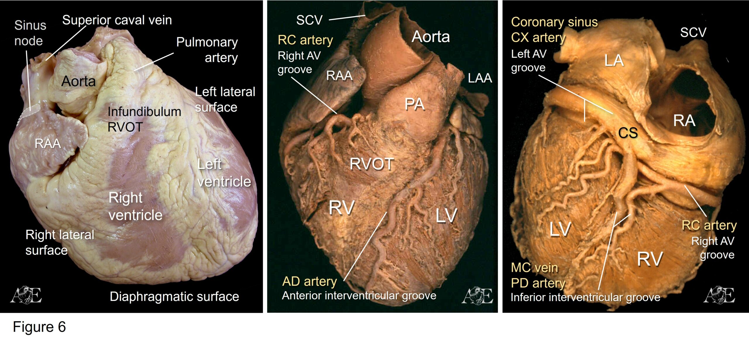
- Anterior surface: This surface is related to the sternum and the chondrocostal joints. It corresponds almost entirely to the right ventricle (RV).In the highest part of this face are observed from right to left:
- Ending from the SCV to the right atrium
- The right atrial appendage (RAA) covering part of the ascending aorta
- The pulmonary trunk artery.
- The apex of the left atrial appendage (LAA). It should be noted here that the right atrium (RA) in an inferior and anterior plane than that of the left atrium (LA).
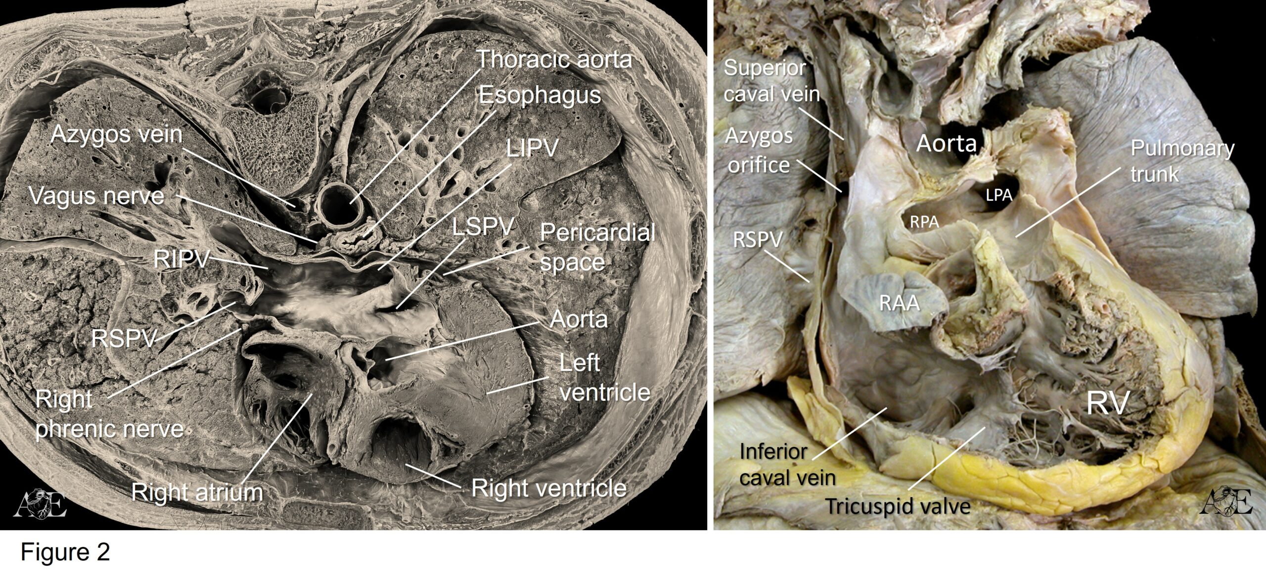
- Right lateral surface: It is composed of the structures related to the RA, such as the SCV. In the middle region of the right lateral surface are located (from the spine to the sternum):
- The body of the RA
- The terminal groove, it is vertical groove that runs from the leading edge of the SCV to the leading edge of the ICV. It corresponds to the terminal crest located in the RA endocardium; and separates the RA from the RAA which projects toward the anterior surface
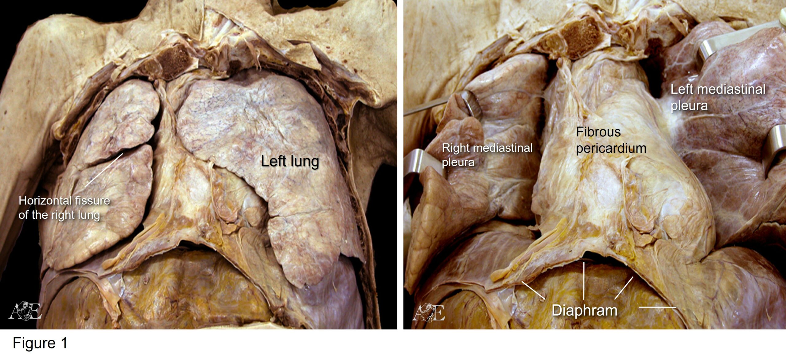
- Posterior to the ending of the SCV the relationships of the right lateral surface are with both right pulmonary veins (superior and inferior) that go to the LA. The right pulmonary artery (at the level of the SCV) and the right main bronchus, and crossing from back to front, the azygos vein.
- Outside the fibrous pericardium, the entire right lateral surface is traveled from top to bottom by the right phrenic nerve (RPN).
- Left lateral surface: composed almost entirely of the LV, upward the ventricle is the implantation base of the LAA in the LA, and posterior and above of the LAA, the left superior (LSPV)and inferior pulmonary veins (LIPV) that open into the LA. The left side of the heart is related to the other mediastinal structures in the following way.
- Above: with the root of the pulmonary trunk.
- Posteriorly: with the left main bronchus, left vagus nerve and thoracic aorta.
- Outside the fibrous pericardium, the entire left lateral surface is relating with the left phrenic nerve (LPN) and pleurae of the lobes upper and lower left lung.
- Inferior or diaphragmatic surface: It owes its name to the fact that the heart is supported on the upper face of the diaphragm. The structures that strictly make up this face are: the LV that represents most of this surface (2/3), and the RV (1/3); and between them the inferior interventricular groove.
- Posterior surface: The main element of this surface is the LA. It is located on a plane higher than LV. On its left side arrive the two left pulmonary veins, superior and inferior. On its right side, but in an anterior plane, is the posterior edge of the RA with the arrival of the superior and inferior cava veins.The posterior surface of the heart relates to:
- Upwards: with the emergence of the right and left branches of the pulmonary artery and even higher up, crossing from right to left, the arch of the aorta that begins to descend.
- Behind the LA is the fibrous pericardium and the esophagus. The relationship with the esophagus is of paramount importance in medical practice and interventional cardiology.
- Superior surface: It corresponds to the emergence of the great vessels. Cardiac rotation and arterial truncus septation causes the pulmonary (PA) and aorta arteries, have their emergence from the opposite side of the ventricle that originates them. Thus, in a more anterior plane, the PA to the left and the aorta in a horizontal plane in a central location, heading to the right. Therefore, the relationship between the two is never parallel but transversal. The aorta is vertical and central; the PA is horizontal anterior and in a plane somewhat higher than the aortic valve.
Grooves and Borders of the heart
Grooves
The fusion of the atria with ventricles is within the plane of the atrioventricular juncions (AV junctions). Also contained within the plane the junctions of the ventricles with the great vessels or arterial trunks, the ventriculoarterial junctions (VA junctions). Because of the major branches of the coronary arteries and the coronary sinus run within these junctions, this zone is also called the coronary groove of the heart.

The AV junctions do not lie in the same plane, because the mitral annulus looks forward and down (highest position of the LA with respect to the LV). The tricuspid annulus looks forward and to the left (because the RA is at the same level as the RV). These rings form two grooves that separate the atria from the ventricles, they are the right and left AV grooves. The right AV groove is traversed by the right coronary artery and the left AV groove by the circumflex artery and by the coronary sinus or great cardiac vein
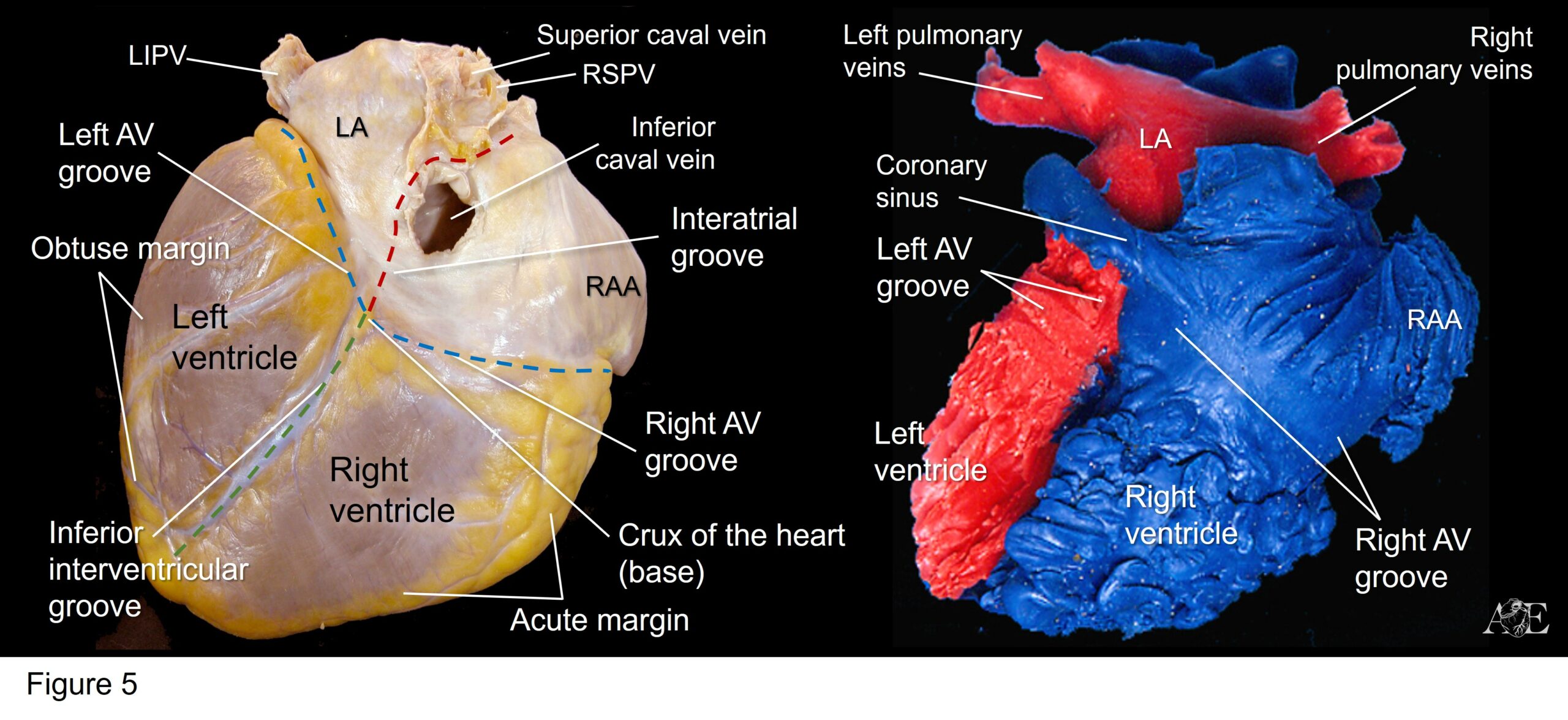
The interventricular septum (IVS) located between the ventricles is oriented on a vertical plane that goes from top to bottom, from back to front, and from right to left. On the anterior surface of the heart is located the anterior interventricular groove. It emerges in the interstitium between the LAA and the root of the pulmonary trunk, reaching the proximity of the apex (corresponding to the LV). This groove is traversed by a branch of the left coronary artery (the anterior descending or anterior interventricular artery) and the great cardiac vein covered by cell-fat tissue. On the diaphragmatic surface we find the inferior interventricular groove. On this surface, the groove runs backwards and somewhat to the left up to the crux of the heart. Like the anterior interventricular groove, it is traversed by a branch of the right or the circumflex coronary artery (inferior descending or inferior interventricular artery) and the middle cardiac vein. . In the inferior or diaphragmatic surface join the grooves of the AV rings and the inferior interventricular groove forms an important crossing point, called as the crux of the heart.
Grooves
The main borders are two, one at the junction of the anterior surface with the diaphragmatic that corresponds to the RV and is called the border or acute margin. This border is sharp when seen from the apex, and is traversed by an homonymous artery (artery of the acute margin) branch of the right coronary artery. At the junction of the left lateral surface with the diaphragmatic, corresponding to the VI, the border or obtuse margin is formed (much more rounded than the acute margin), which is covered by another homonymous artery branch of the circumflex artery.

Media