Iatrogenic biatrial flutter. The role of the Bachmann’s bundle
Emilio Constán, Francisco García, Antonio Linde. Complejo Hospitalario de Jaén, Jaén. Spain
A 63-year-old male patient with a history of atrial fibrillation and common flutter ablated successfully in 2012. Perimitral flutter episode in 2019, performing an anterior mitral line without cessation of the flutter, requiring electrical cardioversion. In 2023 an atypical flutter recurs (Figure 1).

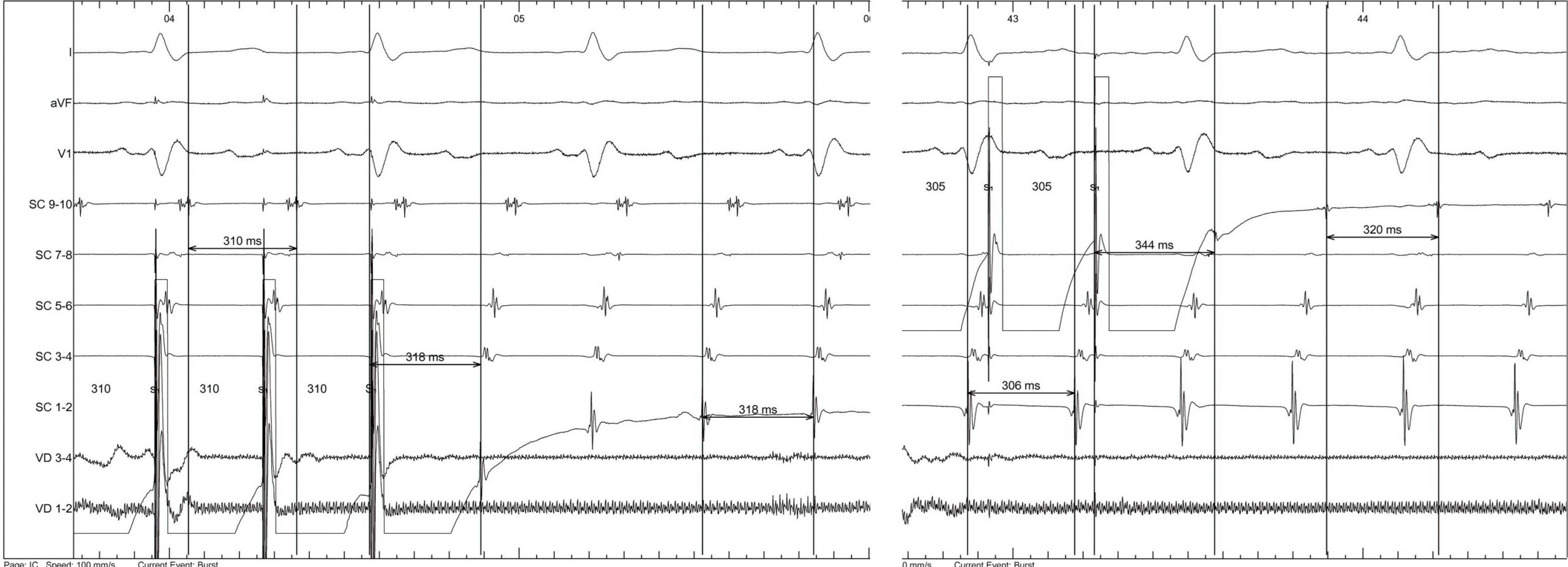
With suspicion of perimitral atrial flutter recurrence, a decapolar catheter is introduced into the coronary sinus, showing exact return cycles at the level of distal dipoles and +24 ms in proximal dipoles. (Figure 2)

Electroanatomical reconstruction of the left atrium was performed with a multipolar catheter, showing isolation of the 4 pulmonary veins and persistence of the anterior mitral line block. Likewise, the activation map of the left atrium revealed the presence of a macroreentrant tachycardia, although without complete cycle integrity around this cavity, for which an electroanatomical map of the right atrium was performed (Figure 3).

Once the activation map of both atria was made, it was possible to appreciate the propagation of the wavefront around the mitral annulus in a clockwise direction, penetrating towards the epicardium through the coronary sinus and ascending through the interatrial septum until the insertion of the superior vena cava, to return again towards the anterior left atrial wall, finally descending through the mitral annulus (Video 1).
Video 1
With the final diagnosis of biatrial flutter with involvement of the Bachmann bundle, radiofrequency applications were made at the anteroseptal level of the left atrium, producing suppression of the tachycardia in the first seconds, remaining non-inducible after the waiting time and reinduction attempts using atrial pacing (Figure 5 and Video 2). No recurrence was observed during follow-up.
Video 2
Although the recurrence of perimitral flutter is greater by performing a lateral mitral line due to the myocardial thickness at this level, the anterior mitral line is the only factor for the appearance of iatrogenic biatrial flutter1. The existence of epicardial connections between the left atrium and the coronary sinus, as well as between the right and left atrium through the Bachmann’s region delimit the biatrial circuit, bypassing the anterior mitral line block (Figures 4-6- Bachmann’s Bundle). Ablation at the level of the interatrial septum allows the termination of flutter2
References
- Mikhaylov E, et al. Biatrial tachycardia following linear anterior wall ablation for the perimitral reentry: incidence and electrophysiological evaluations. J Cardiovasc Electrophysiol, 26 (2015), pp. 28-35
- Nayak HM, Aziz ZA, Kwasnik A, et al. Indirect and Direct Evidence for 3-D Activation During Left Atrial Flutter: Anatomy of Epicardial Bridging. JACC Clin Electrophysiol 6 (2020): 1812-182
Anatomical correlates
By Cabrera & Sánchez-Quintana

-
Bachmann’s bundle (BB) is an anatomical structure connecting the right and left atrial wall and is well considered as the most prominent inteartrial bridge and accounts for the main route of inter-atrial conduction. Regarding its structure, it is a muscular band composed in human of normal (non-specialized) myocardial fibers of the parallel-aligned orientation that cross at the subepicardial level from the beginning in front of the superior vena cava towards the anterior surface of the left atrium where the bundle fuses with the epicardial musculature of the left atrium. (Figures 4,5). (RAA-right atrial appendage; RIPV- right inferior pulmonary vein; RSPV-right superior pulmonary vein; LAA-left atrial appendage; LSPV-left superior pulmonary vein; SCV-superior caval vein)

-
The broad muscular interatrial BB comprising of parallel aligned myocardial strands runs anteriorly relative to the plane of the atrioventricular junction and is visible in the epicardial aspect of the atria under the aortic root when the thin transverse sinus of the pericardium is removed.
-
Although it can be distinguished as a discrete bundle at the anterior septal raphe, it blends in with adjacent musculature of the atrial walls. Its width and prominence varies from heart to heart with median measurements of 4 mm in thickness and 9 mm in height. It has bifurcating branches to both right and left atriums that encircle the atrial appendages (Figure 6).
-
The rightward extension arises in the region of the right superior cavoatrial junction close to the site of the sinus nodal region and the superior extension of the terminal crest or crista terminalis.

-
The leftward extension of the BB bifurcates and encircles the left appendage and then reunite to form a broad band that runs circumferentially around the vestibular wall of the posterior left atrial surface to enter the posterior septal raphe. The superior división continues laterally and traverses the infolding of the atrial wall (left lateral ridge) to pass in front of the orifices of the left pulmonary veins.


