Aortic sinuses of Valsalva as anatomic vantage point to ablate PVC from the LV summit: the role of intracardiac echocardiography
Pablo J Sánchez-Millán, Guillermo Gutiérrez Ballesteros, Miguel Álvarez. Hospital Universitario Virgen de las Nieves, Granada. Spain.
45-year-old male without previous structural heart disease presented with frequent symptomatic drug-refractory premature ventricular complexes (PVC) and mild left ventricular dysfunction related to PVC burden. The suspected site of origin according to the morphology of the ECG is the left ventricular summit (Figure 1).

The anatomic 3D reconstruction of the heart chambers was made using intracardiac echocardiography (ICE) and integrated in the navigation system using CARTOSOUND® module. A 3D-ICE map was performed, allowing visualization of the heart base/fibrous skeleton of the heart and rest of anatomic structures (Video 1).
Video 1
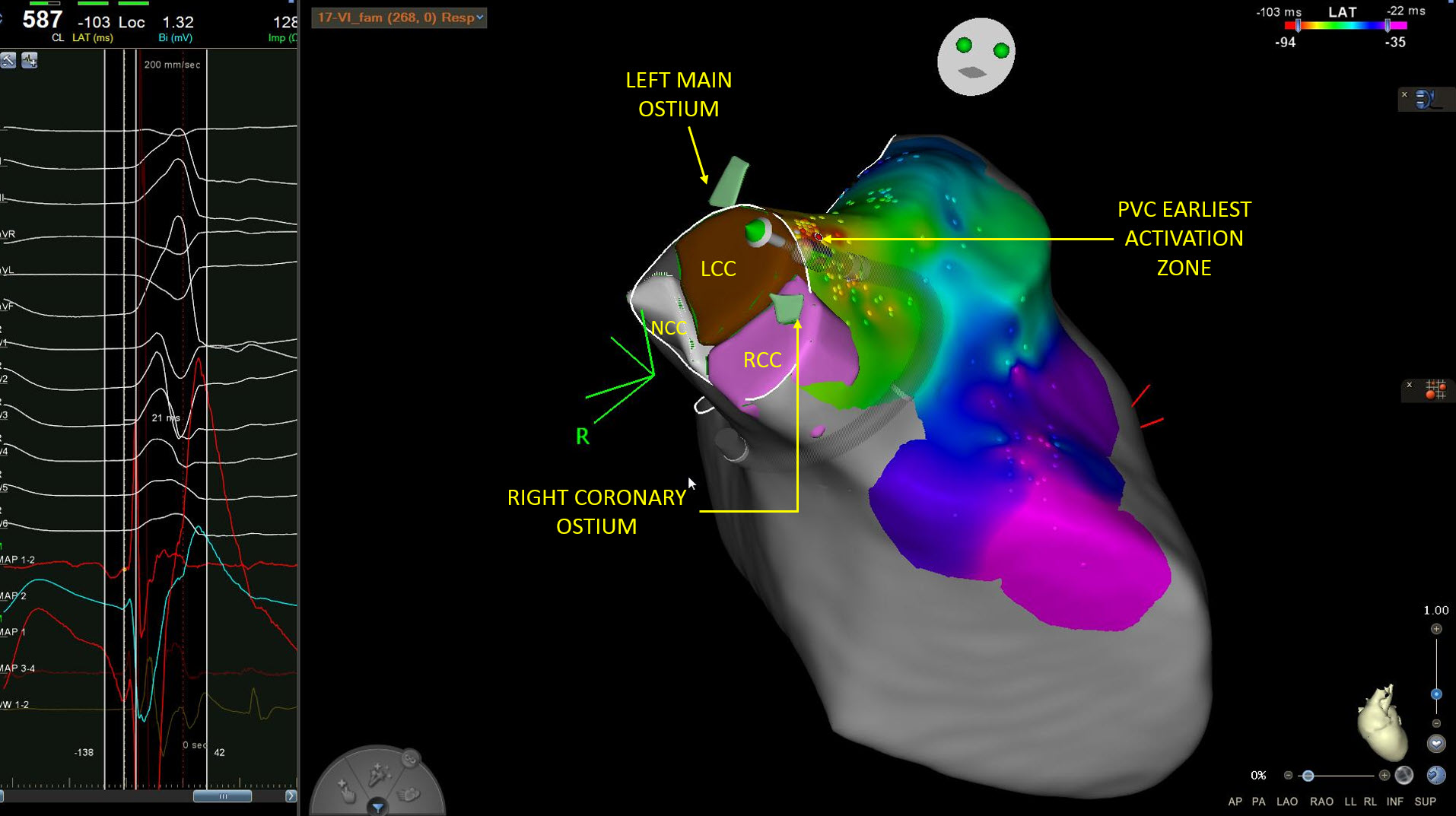
After mapping the right ventricular outflow tract (RVOT), left ventricular outflow tract (LVOT) and distal coronary sinus, the earliest activation site was found in the aortic sinuses of Valsalva or aortic cusps, specifically in the junction between the left and right sinus of Valsalva, also called the left coronary cusp (LCC) and the right coronary cusp (RCC) (Figure 2).

The ostium of coronary arteries, with their anatomic outlets of their respective sinuses of Valsalva, were delineated with ICE and integrated in the map. Without using fluoroscopy, a safety distance to the ostium of the coronary arteries was confirmed (Figure 3) and radiofrequency (RF) ablation was performed between the junction of LCC and RCC (Video 2).

Video 2
The application at that point suppressed the PVC focus with no acute recurrence after a waiting period and during patient follow up (Figure 4)

Reference
Sánchez-Millán PJ, Gutiérrez-Ballesteros G et al. Ablation with zero-fluoroscopy of premature ventricular complexes from aortic sinus cusps: A single-center experience. J Arrhythm. 2021 Oct 3;37(6):1497-1505
Anatomical correlates
By Cabrera & Sánchez-Quintana

-
The left ventricular summit (LVS) is a triangular area located at the most superior portion of the left epicardial ventricular region, bounded by the two branches of the left coronary artery: the left anterior descending (LAD) and the left circumflex artery (LCX). This region was initially highlighted by the group of Yamada and cols as the source of intramural ventricular arrythmias with a high level of difficulty for catheter ablation (Figures 5-6).

-
Ablation within the LVS area may be challenging due to the proximity of the important surrounding anatomical structures such as coronary vessels and the overlying thick layer of epicardial fat that may complicate the approach (Figures 7-8). The anatomical relationships with surrounding structures determines the approach routes for arrhythmias originating from the LVS (green circles).

-
Direct epicardial approach in the inferior area to the great cardiac vein (GCV) (also named the accessible area), where the interventions are relatively safe or may be ablated within the great cardiac vein (GCV) and the anterior interventricular cardiac vein (AIV).
-
The right ventricular outflow tract (RVOT) and the annulus of the pulmonary valve and pulmonary artery (PA) correlates with the lower right sided or septal portion of the LVS and the mitro-aortic continuity to the left sided (Figure 7).

-
The left atrial appendage can cover a significant portion of the LVS area based on its anatomical characteristics
-
The superior vertex of this triangular region is related with the left aortic sinus of Valsalva and the left-right interleaflet fibrous trigone (red arrow, Figure 6) covered by the epicardial adipose tissue and pulmonary trunk (Figure 8). The distance to the semilunar hingeline of the left coronary cusp (LCC) range from 10 to 25 mm.
-
Ablation assisted with ICE imaging allows indentifying anatomical structures of the LVS region that are key to a safer and effective catheter procedure


